


Microscopic discectomy / decompression
Spine surgery
Spine (neck · lower back) · 허리 (요추)
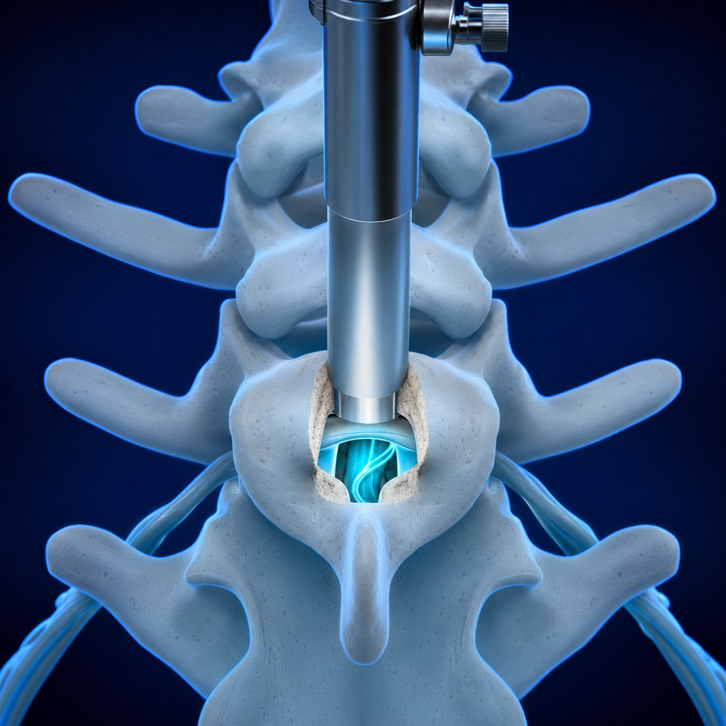
Looking directly inside the spine under a microscope, the herniated disc is removed or the narrowed canal widened to free the nerve. It is the most standardised of spine operations, and complex lesions that an endoscope struggles to reach can be handled within a single field of view.
Anesthesia, hospital stay and recovery time depend on the extent of injury and your general condition. The operating surgeon explains this after the consultation.
When surgery is indicated
- No benefit from conservative or endoscopic treatment
- Refractory or recurrent disc herniation
- A complex herniation accompanied by spinal stenosis
Microscopic discectomy / decompression — benefits
- The incision is kept to a minimum, so bleeding is slight and almost no scar remains.
- The microscope secures the field of view, allowing the procedure to be carried out safely.
- Muscle damage and complications are both minimal.
Process
How surgery proceeds
This is the shared pathway for the Surgery Center. Details for each procedure are covered in the consultation.
- 01
Precise diagnosis
MRI and CT are taken the same day and read by a radiologist that same day.
- 02
Surgical consultation
The operating surgeon explains in person why surgery is needed, what the alternatives are and how recovery is expected to go.
- 03
Surgery · inpatient care
The operation is performed in the annex theatre, and recovery is managed on the ward as the condition requires.
- 04
Rehabilitation · follow-up
After discharge the same team continues to follow your progress and rehabilitation in the outpatient clinic.